“Here is the place where the dead enlighten the living”
The purpose of this lab is to give the medical students a comprehensive knowledge of the morphology of the human body by identifying the structures on the cadavers and to understand its development along with recognition of anomalies which can have clinical implications. Medical students at COMHS attend dissection during their second year at the medical school, and then revisit the dissection hall during their later years to consolidate their learning.
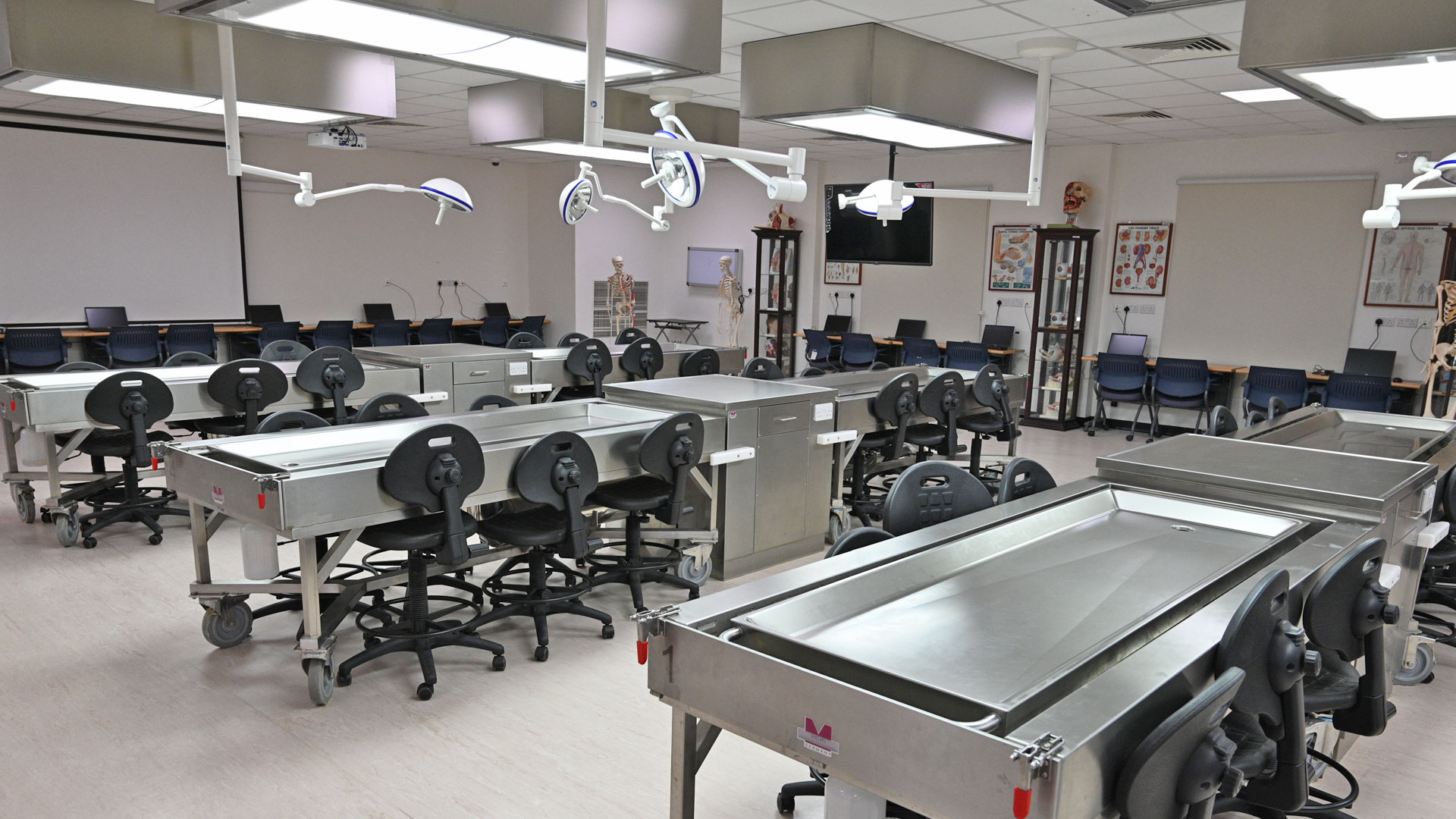
The department of anatomy has “state of the art” gross anatomy laboratory which holds over 100 students at a time. The laboratory has purpose-built stainless-steel tables for placing the cadavers during teaching sessions. This anatomy laboratory is one among the best in the region and has world class facilities for instruction with a full range of anatomical resources such as dissected cadavers, prosected specimen, models, bones, potted specimens and the state-of-the-art technology assisted visual displays.
The lab is also equipped with original bones, skull, radiographs, CT scans and MRI of all parts of the body. For the Neurobiology course, sagittal and coronal sections of the human brain and spinal cord are used. The embryology models in the lab help the learners understand the three-dimensional view of development of various organ/systems that represent the different phases of human growth and development.
The main table in the center is equipped with a high-resolution video camera attached to 8 LED TV screens. This technological enhancement aids in engaging students in the teaching and learning process. The facility enables the instructors to give live demonstration on cadavers to all the students at the same time with the elements of active learning embedded during instructional delivery.
Histology is taught in a digital format using virtual microscopy and the department is in the process of developing a histology webpage exclusively for the use of COMHS students.